Sunday, December 16, 2012

Shape-Memory Polymer Scaffolds

Researchers at Harvard University have recently come up with a method to create polymer scaffolding that can transport drugs and stem cells but doesn't have to be surgically implanted. Instead, it can be injected through a syringe and emerge in the body in the original shape it had because of shape-memory properties. They tested it using scaffolds in circle, heart, and star shapes, and found that it worked each time.

The actual chemistry behind the process of creating these scaffolds relates quite a bit to what was learned in class. The polymers undergo what is known as cryogelation, depicted in the figure below. First the polymer solution is prepared in the liquid phase. Since it is a solution, as we learned in class, the freezing point is going to undergo a depression so the mixture has to be frozen at a subzero temperature. Once frozen, polymerization occurs and ice crystallizes. Then the cryogel is left at room temperature, where it thaws and leaves behind a system with interconnected pores.


Note, however, that this polymerization only occurs in the first place due to the presence of intermolecular forces. The polymer being used is alginate, whose structure is shown below. Because of the hydroxyl groups, there are many polar oyxgen-hydrogen bonds, and the presence of ionized carboxyl groups means that hydrogen bonding will occur. This is what allows the polymers to cross-link so well and form the pores in the first place.


Without the fundamental concepts of intermolecular forces and solution composition as covered in class, these polymer scaffoldings would not even have been possible to make.

Tuesday, December 4, 2012

New Method to Revive Old Stem Cells

Stem cells are cells that are able to transform into cells of other types. This is particularly useful when trying to restore a part of the body internally, as they can continually divide in order to repair other cells. As stem cells grow older, however, their capability to maintain tissues declines. A new method was recently discovered that changes the stem cells so they behave more like younger stem cells, increasing their functioning ability. In particular, this method can help develop cardiac patches for those patients with damaged hearts from their own stem cells. This new procedure is specifically useful as the age of the patient will not affect the success of the stem cell restoration. 

However, in some stem cell therapies, such as those involving bone marrow, there is always a risk of patient rejection. As we discussed in one of our Diigo posts, scientists often use cells from a patient's own body to resolve this issue. We believe that this would also be helpful as patients would not have to wait around for another individual's organ or tissue donations; rather, they would have immediate access, as cells are from their own body.
However, for older patients, the stem cells have become aged and function less than younger cells. Using this new technique, the stem cells from elderly patients can be utilized without being rejected.

This method was created by Milica Radisic and Dr. Ren-Ke Li, two researchers. They created conditions similar to the aged stem cells in tissue cultures, referred to a "micro-environment." In this micro-environment, which the heart tissue would grow in using stem cells from elderly patients. Various factors causing cell proliferation , or the increase in cell numbers, are added to the cell cultures. These infusions caused aging factors to be reduced and cells to be restored to younger versions. In particular, molecules such as p16 and RGN were infusions that caused this to occur. 


The molecule "p16" is a gene that is inactivated when cancers are present in humans. Similar to our topics in biology last year, genes can be inactivated or activated to cease or continue a function. In this case, c
ells are usually limited in their growth through the activation of p16, but when inactivated, p16 actually causes tumors to form. Tumor suppression occurs through protein-protein interactions with p16. Specifically, p16 binds to CDK4, another gene, which causes it to be active.

http://www.chemicalbook.com/CAS%5CGIF%5C189064-08-2.gif
The structure of p16

The bonding that takes place between p16 and CDK4 is of Intermolecular Forces (IMFs). In particular, when CDK4 bonds with other genes, hydrogen bonding networks are created. As we have discussed in class, hydrogen bonds are attractive forces between a hydrogen and an electronegative molecule., usually of a nitrogen, oxygen, or fluorine atom. The picture above shows the large number of hydrogen atoms in the structure of p16. Almost all of these hydrogens are attached to an N or an O, two atoms that are also abundant in p16's structure. This shows that hydrogen bonds exist in p16. P16 is not very structurally stable, particularly due to its "highly dynamic structure as measured by ANS-binding, NMR hydrogen-deuterium exchange, and fluorescence." Because of its weak structure, p16 also self associates and forms dimers. The Kd value of the molecule is only 270 microM for p16. Kd is the equilibrium dissociation constant of products over reactants in the reaction A + B ↔ AB. As discussed last year, it is a specific type of equilibrium constant, K, and is the reciprocal of Ka, the equilibrium association constant. 

In the future, this method can be utilized in many applications. When the stem cells in elderly patients start decreasing in function, they will be revived using this "fountain of youth" technique. Damage caused by heart attacks will be repaired, and defects such as aneurysms can be fixed as well. Although this discovery can be very useful, it might also have several drawbacks. There has always been debate over the moral issues of stem cells. With this discovery, scientists would be tampering with the naturally occurring chemicals, bonding, and genes in the body. Perhaps, for many people, this new method would not be a chemical wonder, but more of a manipulation of nature. 

http://www.newstrackindia.com/newsdetails/2012/11/28/373--Fountain-of-youth-technique-may-help-create-heart-patches-from-old-cells.html


Silk Hearts

Tying in with what we've learned in class, this article from Science Daily explains how silk from a Tasar Silkworm can be used as a scaffold for heart tissue. In other words, how they can use this silk to replace damaged heart cells that are unable to regenerate. They are hoping that this artificial tissue can restore total cardiac function in humans.

Penny-sized silk discs used for heart scaffolding.
These types of studies are so important because, throughout evolution, the heart has lost all regenerative abilities. So when people have heart attacks, all of the cardiac cells that die are lost and cannot be replaced by the heart itself. These studies are conducted to test different types of materials that can patch these dead cardiac muscles. One of their main issues is trying to reconstruct this three-dimensional structure.

Various other materials have failed to work because they were too brittle, rejected by the immune system, or did not allow the muscle cells to adhere with the fibers. Fortunately, researchers are starting to believe that this silk can be a viable material for this heart operation.

The silk has a protein structure that is able to adhere strongly to the heart muscle cells. The coarser material allows the cells to grow and form three-dimensional structures. The re-patched rat heart was able to beat as if it were healthy after being patched with the silk. This is certainly a promising sign for the future and is an indicator that silk can be very successful.


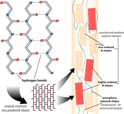
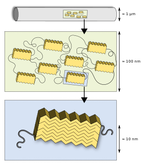
 As the figure to the right demonstrates, the structure of the silk in the Tasar silkworm is very similar to that of spider silk as discussed in a previous blog post, which is what lends it its adhesive properties.

The silk itself is composed of highly crystalline B-sheets crosslinked with less-ordered B-sheets. As discussed in class, the crystalline structure of the more ordered B-sheets allows the atoms to be more tightly connected in a pattern, whereas the less-ordered sheets have an amorphous structure. Furthermore, the pleated B-sheets exhibit hydrogen bonding. As we also learned in class, this is the strongest intermolecular force and results in the interlocking strands. It is because of these properties that the silk is so strong and so capable of adhering tightly to the human body.

Unfortunately, they have been unable to obtain the necessary amount of cardiac cells for the starting material in humans. All tests were conducted on rats and were generally successful. On humans, however, much work still needs to be done. Scientists are in the process of using stem cells in place of these cardiac cells, but trials are still in the early stages.

Tissue engineering is a very important field for improving the overall life of humans. In my opinion, the studies conducted to improve the heart are some of the more important due to the vital role that the heart plays, as well as the fact that heart issues are very common. Any way that scientists can help this problem would be very beneficial to all that suffer from heart conditions.

Wednesday, November 21, 2012

Injectable Shear Thinning Hydrogels

Hydrogels provide a promising new way to deliver drugs or other materials with which to regenerate damage tissue because of their flexibility and biocompatibility. Up until now, though, these could not be fully used because their solid structure tended to degrade under the harsh conditions of the human body, particularly our high body temperatures. To solve this problem, MIT researchers synthesized a special kind of shear thinning hydrogel.

Shear thinning hydrogels are notable because they can switch between a solid and liquid state depending on their surroundings. This makes them ideal as injectable gels because they're liquid when being pushed through a needle but become solid once they're inside the body. However, if the researchers left these gels as is, they would still be subject to stresses of a mechanical nature inside the body and might revert back to liquid.

To fix this, they created a self-healing, reinforcing network inside the gel that only activates when the temperature of its surroundings are body temperature. That way, their new structure would not interfere with the gel's functionality in the needle but would still be able to solve their problem of durability once inside the body.

They opted to create protein hydrogels instead of the more standard polyethylene glycol gels so that they would have better biological properties, and altered the structure so that they would have the desired characteristics. The proteins were loosely held together by links called coils to result in a rope-like structure, and then a second network was built inside that.

At higher temperatures, cross-linking results in a reinforcing network.

At low temperatures, the protein gels are hydrophilic and thus float freely within the gel. As soon as the temperature becomes high enough, though, the gel becomes hydrophobic and groups together, making polymers at the ends of the protein bind to each other. This results in cross-linking so that the gel is significantly more elastic and durable, able to withstand mechanical strength.

Monday, November 19, 2012

Painless Medical Tape

A research team at the Brigham and Women's (BWH) hospital recently created a new type of medical tape that can be removed from skin without pain. Medical tapes before this invention were very secure in terms of adhesion properties, but they damaged skin when being peeled. Current medical tapes have a lot of backing and support that allows it to stick strongly on a patient. However, when taken off, the tape used leaves some of the adhesive material on the skin. Although this does not affect all skin types, neonate skin, which is newer and more recently grown, does face more serious damage. 

Painless medical tape
The new medical tape created has three-layers which retains the strong adhesion properties of previous tape, but also adds a feature that allows the tape to be easily peeled off. There is one layer of tape that serves as the adhesive layer and another layer that serves as the backing support. A final layer exists in between these two, referred to as the anisotropic adhesive interface. The anisotropic feature means that the layer's physical properties depend on direction, opposed to an isotropic feature, which would refer to the properties of materials that are the same in all direction. For example, wood has anisotropic properties, containing lines that go in one direction. The wood contains more strength when the lines all go in one direction, causing wood to have anisotropic properties. 

Anisotropic features have advantages, being stronger in a certain direction. The scientists from BWH used laser etching and a release liner in order to make the anisotropic layer of the medical tape. This feature allows the medical tape to have a much higher shear strength as it is now dependent on direction. The release liner allows for the medical tape to be removed with little force. Solving the previous issue, if any adhesive remains of the skin, it can be gently rolled off using a finger. In other words, the medical tape would come off skin more easily. Release liners work by using silicone releasing agents, which have a low surface tension of wetting. Wetting refers to the characteristic of how a liquid deposited on another liquid or on a solid spreads out. Because silicone, a key component of release liners, has a low surface tension energy, it does not spread too much when on another layer of the medical tape and, ultimately, the skin. Thus, it can be pulled off easily without any damage to the skin. Frequently in the past, carbon-carbon backbones were utilized instead of silicone polymers, which caused a more rigid structure and, as a result, harm to skin. Silicone polymers, however, have methyl groups which interact only slightly with one another, producing little surface tension energy.  Additionally, silicone has a low adhesion in terms of sticking to the skin. When materials have diffusive adhesion, the molecules of each are soluble in the other material. They move around, and when joined, connect through diffusion. In particular, polymer chains demonstrate diffusive adhesion effectively, in which the ends of one molecule diffuses with another. 


Over 1.5 million injuries occur in the United States due to medical tape removal. Using this medical tape, this number can be reduced greatly, affecting numerous patients positively. 


http://articles.timesofindia.indiatimes.com/2012-10-30/health/34816599_1_tape-skin-release-liner 

Sunday, November 18, 2012

Walking Bio-Bots

For the first time, researchers in the University of Illinois have created an autonomous robot made from plastic and living cells. With just hydrogel, heart cells, and a 3D printer, these scientists have created miniature "bio-bots" that are capable of walking by themselves, without the use of electricity.

The Bio-Bot is 7 mm long and can move on its own
Each bot has one long, flexible leg that rests on a short, stout leg for support. The long leg is covered with rat cardiac cells. It pulses like a heart and as it beats, the bio-bot propels forward. The rhythmic expansions and contractions pull the little robot forward in what looks like a swimming motion.

The hydrogel part of the robot was made by a 3D printer. This makes the production of robots easier because more shapes can be made faster and easier. Because it is easy to print these plastics, several changes can be made fairly easily. The researchers can use trial and error as much as they need to improve the design of the bio-bot.

The bio-bot's unique structure allows it to manipulate into different shapes that would not be possible if it were made of metal and hard plastics. The soft plastic and cells allow the robot to be flexible, as well as function without the use of electricity. Rashid Bashir, one of the head engineers, stated “As engineers, we’ve always built things with hard materials, materials that are very predictable. Yet there are a lot of applications where nature solves a problem in such an elegant way. Can we replicate some of that if we can understand how to put things together with cells?” So far, they have been successful in creating functioning robots.

The researchers hope that these bio-biots could be used to detect chemicals in water, climb walls, or function as a sensor to react to certain elements in the water. If the bio-bots can be made using cells that respond to different stimuli, like chemical gradients, then they can definitely be used in drug screening and chemical analysis. The way the bot's motion changes would indicate the reaction of the cells to the environment and whether or not a similar reaction may occur in humans. It is the researchers' hope that if they can get the bot to move toward chemical gradients, they could design something that can look for a specific toxin and attempt to neutralize it.

A depiction of the movement of the robot
The scientists plan to continue working on this discovery. They hope to improve the bots to develop control and function. For example, they plan to create robots that can respond to light, climb steps, and utilize different numbers of legs. They believe this is the first step of many in this exciting field that utilizes nature for beneficial purposes and functions.


http://news.discovery.com/tech/walking-bio-bot-made-with-cells-gels-121115.html
http://news.illinois.edu/news/12/1115bio-bots_RashidBashir.html


Growing organs from your own cells

Andemariam Teklesenbet Beyene had a cancer-ridden windpipe, but thanks to some brilliant scientists, he left with a unique windpipe, one that was created from his own cells. This was the first time that a synthetic organ was successfully created and used to cure a person. The success marks the beginning of a time when organ transplants will no longer require a waiting list or debilitating drugs.

The process for creating an organ capable of functioning in the human body with everything else that is going on is quite complicated with trial and errors. There are many factors that must be in check for the organ to be produced properly. An amount of the patient's cells must be extracted and nurtured, than factors such as the shape, temperature, pH, and others must be monitored to ensure the organ grows correctly. Also, the organ was tested in a bioreactor, which mimics the conditions in the human body. It was only after all this that Beyene had his synthetic windpipe transplanted, and months later Christopher Lyles, had the same treatment for cancer. Due to the complexity of the process, only simple organs like windpipes are able to be constructed, while more complex organs like hearts are still impossible.


http://www.bbc.com/future/story/20120223-will-we-ever-create-organs/1


Saturday, November 10, 2012

Bulletproof Spider Silk Skin

Under Project 2.6g 329m/s, researchers worked in collaboration with the Forensic Genomics Consortium Netherlands to create one of the most creative and shocking tissue engineering projects: bulletproof skin made of spider silk.

Silk has always been known to be among the strongest natural protein fibers in existence, but spider silk in particular has remarkable properties, its tensile strength larger than that of steel and its flexibility enough to be woven into a web. More intriguingly, spider silk has been proven to be biocompatible, raising the question: can spider silk replace the protein keratine, which is currently used to provide strength in human skin?

Project 2.6g 329m/s answered this question by growing a patch of human skin that consisted of a spider silk matrix in between the dermis and epidermis. As the video below attests to, this artificial skin was even able to stop a bullet.


But what exactly about spider silk makes it so flexible and strong? The answer to this question lies in its chemical structure. Similar to Kevlar, spider silk is a combination of crystalline structures in an amorphous matrix. Its main protein is called fibroin, consisting primarily of the amino acids alanine and glycine. Fibroin has two differently structured regions that contribute to the tensile and elastic properties of silk.

Hydrogen-bonded polyalanine regions.
The first are the polyalanine regions, which give silk its strength. These are exactly what they sound like: repeating blocks of multiple alanine molecules linked together. Due to hydrogen bonding, these different blocks have strong interactions with each other and connect together in a highly ordered crystalline structure. Cross-linking individual proteins in this manner is what results in such high strength.

In the glycine-rich spiral regions, a sequence of five amino acids is repeated over and over similar to the polyalanine regions, but the twist is that the molecule takes a 180 degree turn at the end of each sequence. This results in, as the name suggests, a spiral which is so densely twisted that some types of spider silk can even stretch up to 4 times its original length.

Crystalline regions juxtaposed with amorphous regions.

Further contributing to the elasticity, the glycine-rich spiral regions comprise the amorphous areas of the silk. Somewhat less ordered polyalanine regions connect the amorphous areas to the more ordered polyalanine regions, resulting in an overall structure that is both immensely strong and surprisingly flexible.

Friday, November 9, 2012

Artificial Blood Vessels Created with Aid of Lightning

Scientists at the Texas A&M University have been looking into a way of building artificial blood vessels, and by this, artificial organs by using lightning bolts. It may be questioned how lightning could possibly help create an object, but, in fact, the high electric discharge from lightning can actually make a series of miniature tunnels or holes in materials. For example, if a block of plastic is electrically charged by gaining or losing electrons, lightning is able to permeate areas all around the plastic block. 

A tap of a nail is being used to produce a Lichtenberg figure.


 In particular, the tunnels created when lightning streaks are applied to plastic blocks are very similar to the features of the capillary system of the body. In the human body, these "tunnels" are actually arteries and vessels. When tunnels are created in this artificial manner, they are quite accurate in the sense that they are the same size as capillaries and are connected internally. Lightning is used in this process because it allows tunnels to be made throughout the block instead of simply at several places on the surface of a block. If made into artificial blood vessels, blood will be able to flow throughout the entire organ. 

In particular, a bolt of lightning allows a similar effect on a plastic block to the result if electrons were trapped inside the plastic block. When electrons travel, they create an electric current, which is what is present in the block. The current does exit the block, causing the plastic to be damaged because of the heat produced by the current. This is what ultimately creates the various tunnels. Scientists replicate this by using a Wimshurst machine or a Van de Graaf generator to create static electricity. This electricity is discharged, causing the plastic to have a pattern of charges. Called a Lichtenberg figure, the ending plastic block with tunnels was originally created in a different process in the eighteenth century. Back in this time, scientist Georg Lichtenberg utilized powdered sulfur and minium to test the process. Powdered sulfur was found to be negatively charged from the friction between it and the container it was set in. Because opposites attract, the negatively charged powdered sulfur attracted to the positive spots on the plastic block surface. The minium, a red powder, was positively charged due to friction as well, and was attracted to the negative spots. This combination allowed hidden features to be revealed in the figures and allowed a better visual to see.

In the past, prolithography, a method used to create computer chips, has been utilized to make these 3D artificial vessels. The technique of using lightning streaks is, in this way, quite efficient as the entire process only spans a couple of seconds. Using this method, artificial vessels are not only less of an expense to make but are also faster. 

However, there is also a potential disadvantage to this process.When each block of plastic is created, the tunnels present inside each are constructed in the same manner, but produce different tunnels. On the other hand, when using prolithography, all the tunnels end up exactly the same. This might be inefficient if certain systems need to be reproduced for various organs. Other scientists argue that artificial organs do not need to be exactly the same. Rather, the organs only need to have several processes in common, such as those to gather nutrients and eliminate waste from the body.

Overall, this is a revolutionary discovery in the world of tissue engineering. Because of this research, scientists will soon be able to approach using implant cells, which can be combined to create vessels, and eventually, organs. The cells would continue growing, and the plastic used would someday degrade, which does not cause harm to the body. However, the process still does involve many risks, and so, it will be implemented in animals after many years. Humans will be tested if the animal test runs successfully. 


http://news.discovery.com/tech/lightning-artificial-blood-vessels.html 
http://www.popsci.com/diy/article/2008-02/trap-lightning-block 
http://205.243.100.155/frames/lichtenbergs.html

Friday, October 26, 2012

Lab Grown Lung Video

The following excerpt from a NOVA program on tissue engineering shows a lab grown lung breathing on its own.


Thursday, October 25, 2012

How to Make Your Own Jellyfish

Jellyfish, one of the strangest organisms known to man. They can be found in every ocean in large quantities, and they are one of the oldest organisms in existence, surviving for nearly 500 million years! Yet, something this old is not the slightest bit wise, as it doesn't even have a brain.

However, the bioengineers at Caltech and Harvard chose to artificially create a jellyfish. You may think why do we need more of something that is already so plentiful, and potentially dangerous. Well, the bioengineers explain that jellyfish have a simple structure to them. Jellyfish are pretty much just "a pulsating bell, a tassel of trailing tentacles and a single digestive opening through which it both eats and excretes." The bell of the jellyfish contains its vitals, and by understanding how it works, the bioengineers began their attempt at creating an artificial jellyfish.

A polymer jellyfish
A schematic of the bell was created, detailing the tissue used. Using the schematic, the bioengineers began constructing the jellyfish. First, they cut a thin polymer membrane, a material similar to jellyfish tissue, into the shape of a jellyfish. Then, they placed rat heart cells and protein into the polymer. The rat hearts served as the vitals, while the protein served as a guide for the rat hearts to grow correctly. Once this was finished, they submerged their invention into a tank and sent a current through it. Amazingly, the invention came to life and had movements similar to a real jellyfish.

This successful experiment can lead to many new discoveries in the future. Despite the simplicity of a jellyfish, this is a great starting point for future construction of more complicated organisms, and eventually to the possibility of being able to operate on humans. In the meantime, the bioengineers at Caltech and Harvard will continue to improve their jellyfish, hoping to give it the ability to move without a current and capture its own food.

http://www.time.com/time/health/article/0,8599,2120119,00.html

The following video sums up the creation of the jellyfish:

Wednesday, October 24, 2012

Mass Production of Tissue- Engineered Meat a Possibility

The CEO of the company "Modern Meadow" has announced that his company has and will continue to work on the process of 3-D bioprinting leather. The company believes that the leather will be mass-produced by 2017 and manufactured meat may follow in several years. Modern Meadow's main focus will be on leather for the time being.

While meat may be a more helpful and useful use of their funding, skin is a much simpler material to imitate. Additionally, leather will be a much less controversial material to the public. The company has found that only 40% of the population would be willing to try this manufactured meat, while very few would have a problem using this mass-produced leather. In fact, this process should go over very well with the public, as the scientists will not need to kill any animals.

The scientists have already come up with the process and just need to find a way to increase the scale of production. In other words, they face engineering, not scientific obstacles. The five step process includes taking punch biopsies of donor animals. While these animals need to be dead, the researchers are using animals that were already going to be killed for meat or skin. The researchers then make necessary modifications to these cells.

By using a bio reactor or growth apparatus, the cells multiply from millions to billions in number. They are then centrifuged to eliminate the growth medium of the cells and to lump them together into clumps of cells.

These aggregates are fused together using a process known as bioassembly. There are still several possible processes and Modern Meadow is still trying to find the best approach.


The complex process of manufacturing leather


The scientists then put these fused cells into a bioreactor and given time to mature. They create an environment which allows "nature to take over" the growing process of these cells. This increases the growth of the collagen cells

Several weeks later, the food supply is cut off. The skin tissues turn into hide. The leather goes through a short tanning process that minimalizes the use of toxic chemicals.

The future of this field is very promising. The researchers are confident of the science involved in this process. They just face engineering problems that they will overcome in the next few years. If the growth is successful on a large scale, thousands of lives of animals would be saved. If meat can be produced in several years, millions of animals’ lives would be saved. Tissue-engineering may be the answer to prevent different types of food shortages around the world.
 
Source:

Tuesday, October 16, 2012

UCLA Discovers Migrating Cells' Tendency to Turn Right

The diagonal pattern formed

It has often been said that observation is the key to success in science. A group of researchers at UCLA brought this concept to a whole new level by keeping a close watch on cells as part of a study. Not only did they find patterns in the migration of the cells but they also were able to spot a certain right and left symmetry.

In particular, the UCLA research team created a culture surface of cells and used various protein substrates that were either cell-repellant and cell-adhesive. The floor of the lab surface consisted of stripes of tile and carpet, which created the overall surface change that cells face in a body. Microtechnology was used to observe the cells.

The difference in the surfaces used brought about a completely unexpected discovery: that cells were able to reorganize themselves based on the surfaces they encountered. However, in addition to simply cross the different types of surfaces, the cells also formed a pattern. These migrating cells turned twenty degrees to the right in parallel rows, which created an overall diagonal pattern.

It was not expected that the cells would form this type of pattern, let alone, a pattern at all. The study had been conducted to observe the process of vascular cells forming structures in cultures. Instead, the researchers had observed a left-right assymetry caused by the surfaces.

In the future, this fact can be utilized to have control over the migration of cells and the shapes formed by these cell cultures. Substrate interfaces can be used to develop cells in different ways. This study adds to knowledge of tissue architecture. In particular, this reserach can allow a person's own stem cells to be used for the development of organs. Becuase there are a limited number of organ donations, this research can help to solve this issue.


Friday, October 12, 2012

New Hydrogel for Tissue Engineering

One of the most important considerations to make in tissue engineering is the kind of material you use, because it has to be both capable of sustaining the constant stress placed on it by the body and still be biocompatible. This can make it very difficult to replace or grow new tissues with synthetic materials because oftentimes something that is strong enough might have too high toxicity levels, and something that's proven to be perfectly safe might simply be inadequate for the job.

Hydrogels can be very stretchy. 

An example of such a material that has been troublesome in the past are hydrogels. As the name suggests, these are composed mostly of liquid yet behave as a solid because of their structure. The liquid molecules are dispersed throughout a solid and are cross-linked in such a way that makes them have a jelly-like consistency.

Because they are both water-based and biocompatible, hydrogels make ideal candidates for tissue engineering applications such as cartilage replacements or usage in spinal disks. However, the problem lies in their weakness. Current hydrogels have been very brittle and thus unsuitable, so many researchers have been attempting to create a hydrogel that would be stretchy and strong enough to suffice.

At last, Harvard researchers have had a break-through. The hydrogel they created is made of two common polymers, polyacrylamide and alginate, combined in an 8:1 ratio, and although the two polymers aren't very impressive on their own, their combination is tough, self-healing, and can be stretched to 21 times its original length without breaking.

The alginate chains bond weakly with one another and trap calcium ions, and when the hydrogel is stretched, the bonds but not the chains break, thus releasing the calcium ions and allowing the gel to expand. However, the stretchiness is increased when the alginate is combined with polyacrylamide because of the grid-like, cross-linking phenomenon that occurs.

The polyacrylamide chains bond very tightly with the alginate chains, so the breaking of bonds diffuses across a wide area rather than being concentrated in one place and risking a crack or tear. The self-healing comes in because the alginate chains are able to re-form the ionic bonds between them, essentially renewing the hydrogel.