Friday, February 15, 2013

Heart Tissue Grown Using Carbon Nanotubes

You have all probably noticed that hydrogels are a huge topic of interest in our blogs. Hydrogels are very important in the field of tissue engineering, especially due to their flexibility. Recently, a hydrogel was created using carbon nanotubes that would serve as a scaffold for cardiac tissue that would "beat spontaneously."

In heart attacks, cardiomyocytes, or muscle cells, are attacked. If artificial heart tissue is made, it must follow the structure and function of the heart. The heart is an organ that transmits signals to cells for the regulation of muscle contractions (heart beating). Heart tissue that is lab-grown must have a scaffold that is electrically conductive so it can also do this. 

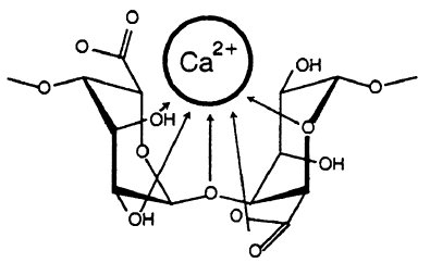
Cardiomyocytes have been shown to grow on alginate or gelatin scaffolds in the past. Alginate is a polysaccharide made up of α-L-guluronic acid and β-D-mannuronic acid. Because alginate is found in seaweeds, its structure is very gel-like and flexible. When alginate interacts with metal ions, such as calcium ions, hydrophilic gels are formed. In terms of structure, both poly guluronic acid and poly mannuronic acid bind to calcium ions. However, poly guluronic acid interacts more strongly to calcium by cross linking, and it also has more hydrogen bonding between the carboxyl groups and OH groups. Alginate structures with a greater amount of poly guluronic acid thus have stronger structures. 

polymannan
Sodium polymannuronic acid


poly guluronic
Sodium polyguluronic acid

However, the combination between polyguluronic acid and calcium does not have a high specific heat, making it a poor conductor and unacceptable for an electrically conductive hydrogel. Another material, gelatin, is also not a very good conductor, but when bonded with methacylate monomers, a more structurally stable molecule is formed.  Methacrylate, C5H8O2, adds more covalent bonds to the overall structure.


The carbon nanotubes had a crosslinked methacrylated gelatin film on it, allowing the hydrogel to be formed. This new hydrogel imitated the Punkinje fibers in the heart that have conduction capabilities. It was also seen that these new cells beat at a faster rate than tissue that contained only gelatin and could use an 85% weaker electric field to beat.

In the future, these nanotube-grown cells will be continually used in cardiac tissue applications, as well as in other organs of the body that require the withstanding of contractions. 

http://cen.acs.org/articles/91/web/2013/02/Carbon-Nanotubes-Help-Grow-Beating.html

Thursday, February 14, 2013

Spray-On Skin Kit

Avita Medical has created a new spray-on skin kit known as the ReCell Kit that does exactly what it sounds like - generates new skin for you after a simple spraying mechanism. It can be used to treat burns, wounds, and even correct blemishes for cosmetic purposes, such as altering abnormally pigmented skin or improving the appearance of scars.

Although this kit does seem to work wonders, its basis lies strictly in the scientific realm. In short, the keratinocytes and melanocytes of the patient are harvested and then suspended in a solution in which they can multiply. It is this solution that is sprayed onto the patient and results in the growth of new skin. Because the harvested cells come directly from the patient, any risks of the immune system rejecting the treatment are eliminated.

The reason these two types of cells are used in the solution is because they are the building blocks of skin. Keratinocytes in particular are of utmost importance because they are responsible for the production of keratin, a structural protein without which much of our bodies would fail to function. One of the most crucial of keratin's properties is its strength even at very high temperatures.

This is because its toughness comes from its supercoiled nature in which the polymer is kept together through cross-linking brought on by both intermolecular forces and covalent bonding, as seen in the diagram. Covalent bonds form between sulfurs to result in disulfide bridges, which kept the protein rigid. However, even at the high temperatures of the body, the bonds stay as they are. Thus, the dissociation of the bonds is nonspontaneous, which means the value of change in Gibbs Free Energy is positive.

Furthermore, since such dissociation would result in more positional arrangements, entropy would increase and thus change in entropy is positive as well. This means that the sign of enthalpy must be positive or else the change in G would be negative, so the bond dissociation is endothermic. Eventually, at a high enough temperature, the bonds will dissociate because of this fact.

Thursday, February 7, 2013

3D Bioprinting Tissue

A new 3D bioprinter has emerged from the University of Toronto with the shocking ability to print skin by making ample use of colligative properties as they relate to solutions. Although this printer is still in its early prototype stages, it has the potential to be used to print skin, organs, and even artificial food as discussed in an earlier blog post.

The printer works much like a normal printer, with seven reservoirs comparable to the color cartridges normally found. The main difference, of course, is that each of these reservoirs contains living cells instead of ink. This part is fairly standard for other 3D bioprinters, but what makes this different from the others is that it doesn't rely on the traditional layer-by-layer assembly often seen, which makes this less time-consuming and more readily available for applications such as burn dressing.

Each of these living cells from the reservoirs are released into a stream of a compound known as sodium alginate, pictured at left. This polymer is a derivative of algae and is thus biocompatible in addition to strong and flexible when in gel form, which makes it ideal for tissue engineering applications. However, sodium alginate is soluble in water, so in this form, the polymer cannot be used to make any tissues or organs. The next steps of the chemical process used in this bioprinter are therefore made to ensure this polymer can be changed into an insoluble form. Otherwise, the cells would do nothing more than merely sit in solution.

To solve this problem, the stream containing the cells and the sodium alginate flows into another reservoir containing calcium chloride. When sodium alginate comes in contact with this solution, most of the sodium ions exchange with the calcium ions to result in calcium alginate. The big difference between these two is that calcium alginate is insoluble. This is because the electrostatic force between the very positively charged calcium ions and the anionic polymer overcomes the hydrogen bonding and other solute-solvent interactions between the water and the alginate. The result is that the calcium ions crosslink the polymer by joining the strands together, and in the process, result in an insoluble gel. This is depicted in the figure below.


Using this gel, the printer can easily spin the result into organs and other tissues.

Thursday, January 31, 2013

Silk Protein Fibroins Combined with Albumin for Tissue Engineering

More unique and efficient biomaterials are being discovered that allow tissue engineering to be successful and progress onward as well. A research team part of the Indian Institute of Technology (IIT) recently found that silk protein fibroin works well with tissue engineering and drug delivery.

The chemical structure of silk protein fibroin is what contributes to its great value in these bioengineering applications. We wrote about silk from spiders and its use to create artificial hearts in a previous blog post. Similarly, silk fibroin is a protein that comes from moth genera, such as spiders. Proteins consist of amino acids (as we learned in biology last year!). Silk consists of, specifically, 60% Glycine, 20% Alanine, and 20% Serine.


Silk Fibroin structure
These IIT researchers utilized a combination of silk fibroin and the protein albumin in order to provide an overall stronger material. Electrostatic interactions exist among the silk fibroin's carboxyl groups and the amino groups of albumin.
Albumin structure
Amino groups, being positively charged, attracts to the negatively charged carboxyl groups, creating a strong combination of electrostatic interactions because opposite charges attract. This strength contributes to the "encapsulation" and "drug retention" of the drug.

These researchers then created fibroin-blended nanoparticles. FITC (Fluoresceine-isothiocyante) nanparticles are not toxic to body cells, which they can reach. When methotrexate, another drug, is added to the fibroin-blended nanoparticles, delayed drug release increases.


Methotrexate structure
Additionally, the nanoparticles are able to remain at a site longer, which may allow them to be used with methotrexate as nanocarriers or hydrophobic therapeutic agents. 


http://nanotechweb.org/cws/article/lab/52196

Sunday, January 27, 2013

Scientists Modify Body pH Levels to Prevent Cancer Spread

I am sure we all know that a definite cure for most cancers have not been found yet. However, researchers are currently looking into ways to prevent and slow the growth of tumor cells. In particular, scientists of the Moffitt Cancer Center and Wayne State University School of Medicine have been researching the effects of acidity, or levels of pH, on cancer cell invasion. In the study conducted, it was determined that increased fermentative metabolism and inefficient flow of blood to tissues contribute to increased pH levels in tumors.

Essentially, immunodeficient mice with tumors were tested. The states of these mice would ensure that they would be incapable of normally surviving these tumors. Using microscopy, the areas of the mice with the lowest pH values were the places in which the tumors were most strongly present. It was found that the tumor invasion and spreading continued in regions of the body with "normal or near normal pH levels." Ordinarily, pH values at biochemical standard state conditions are defined to be at 7. As we learned in class, the Gibbs Free energy (G) depends upon numerous factors. One of the formulas to calculate G is G = Go + RTlnQ, Q being the reaction quotient. Because the Q directly relates to the concentrations of the various compounds reacting and forming in the body, it is evident that a low Q must be the result of greater concentrations of reactants. In order to take account for this, the researchers used sodium bicarbonate to neutralize the acid and bring more stability. This compound, NaCHO3, is known as an amphoteric, a molecule that can react with bases and acids. Researchers found that addition of sodium bicarbonate stopped the cancer invasion.

Scientists have figured out that increased glucose metabolism is a cause of this higher acidity. It causes an "abnormal vascular network" that disrupts blood from being delivered, along with oxygen. This in turn, causes less oxygen and more glucose fermentation in areas with tumors. Researchers have labeled this process to be called "niche engineering." Tissue cells carry out niche engineering that causes increased acidity, which does not prove fatal to malignant cells but can increase malignant cell spread and invasion. In the future, scientists plan to focus on this tissue "niche engineering" with buffers and other chemicals that can increase acidity to balance out the decreased pH levels.

http://www.sciencedaily.com/releases/2013/01/130125111145.htm

Saturday, January 19, 2013

Collagen Scaffolds: Slice, Stack, and Roll

Tufts University researchers have invented a new method for creating collagen scaffolds officially called bioskiving and colloquially referred to as "slice, stack, and roll" that would enable it to be more useful than in its normal form. Collagen is a protein present in the flesh and connective tissue that is often utilized for tissue engineering purposes because of its strength and biocompatibility. However, it can be inefficient because production techniques often lower its natural strength.

In bioskiving, the first step is to decellularize cut sections of collagen with a detergent, which leaves behind a matrix of bundled collagen nanofibers without any excess. Next, the sections are sliced into very thin sheets and stacked in such a way that the alignment of the fibers alternates throughout the stacks in order to increase the tensile strength. Finally, the stacks are rolled around Teflon-coated glass rods to maintain the fiber structure, which is important for the strength of the overall material.

A picture of this process is shown below:

Collagen's strength is due to its chemical structure, which consists of three polypeptide chains, each of which are in the shape of a helix. This strength is further compounded by the fact that the amino acids in this protein are very small, allowing each chain to be twisted together tightly. In the chemical structure, it can be seen that hydrogen bonding is present because of the electronegative oxygen and nitrogen. This helps keep the strands tightly together.

There are also cross-links between the three-stranded collagen molecules, which lends the protein high tensile strength. This means it is very resistant to breaking apart under pulling forces, while still retaining some measure of elasticity. Because of all these different types of strength, collagen remains the ideal protein to use for such application, and the slice-stack-roll process was able to retain these properties.

In a somewhat different vein, collagen has one other important property: its relative solubility in water. As seen in the structure, collagen is a very polar molecule, with the hydroxide groups and electronegative elements. Since like dissolves like, it is not hard to make solutions with collagen in water, which makes it a good choice for biological applications. If collagen was insoluble, it would not be suited for this work.

Wednesday, January 16, 2013

Stem Cells and Tissue Engineering


Because a lot of our twitter and diigo posts mention stem cells, I thought it would be a good idea to make a blog post about it.

A lot of good information comes from here:
http://stemcells.nih.gov/info/basics/basics1.asp
But in summary: 
Stem cells are found in nearly every multi-cellular organism. They are special because they have the ability to renew themselves through mitotic cell division. They can then differentiate into many types of specialized cells. So a stem cell can change into a lung cell, brain cell, heart cell, etc. They can become tissue or organ-specific cells with special functions.

Stem cells are very important to living organisms. In the blastocyte, the 3- to 5-day-old embryo, the inner cells give rise to the organism's entire body. This includes all of the specialized cell types and organs like the heart, lung, skin, sperm and eggs. In adult tissues like bone marrow, brain, and muscle tissue, populations of adult stem cells have the ability to generate replacements for cells that are lost for any reason, like normal wear and tear or disease.
A diagram of Stem Cells and their modified forms

Now scientists are working to use stem cells for therapy and rebuilding of organisms. There is a lot of work to do, but it is a very popular field, so a lot of work is going into it. There is a bright future in the field of stem cells.

These stem cells are able to generate crucial body parts like hearts, eyes, lungs, and brains. If they can be perfectly understood and used often, then several diseases can be cured. You can read the rest of the blog posts or tweets to see some more examples.

There is so much more to stem cells than I can include in this blog post, plus several studies conducted using them. There's no doubt that future tissue engineering news will include stem cells. 

Of course there is the famous controversy behind it involving the use of fetuses to gather stem cells. It appears to be the biggest setback to this field. However, there is no denying that the future of stem cells is bright